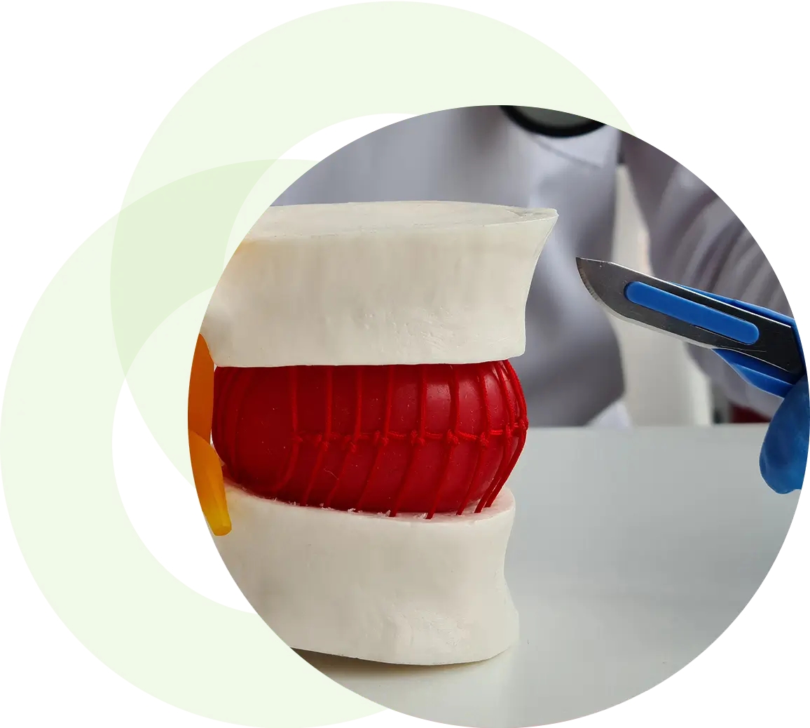
Percutaneous Discectomy
A percutaneous discectomy is a minimally invasive procedure used to treat damaged spinal discs.
If a disc herniates or ruptures, the inner gel material leaks out of the disc into the spinal canal. Once the material makes contact with spinal structures such as nerves or muscles, it causes pain, irritation, and inflammation. The goal of a percutaneous discectomy is to eliminate pain by shrinking or removing the material surrounding the injured disc.
Frequently Asked Questions
The surgeon uses a medical imaging technique known as fluoroscopy to identify the problematic disc. Rather than making an incision in the skin, the surgeon uses fluoroscopic imaging to insert a needle into the disc. Using heat waves, the surgeon then removes the damaged disc material, easing pain and relieving pressure on compressed nerves.
A percutaneous discectomy can be performed using local anesthetic, which reduces the chance of complications.
A percutaneous discectomy can help manage pain associated with herniated discs and related nerve issues such as sciatica. Nerve pain can be debilitating, and it can affect a patient’s quality of life – if your pain doesn’t cease after six months or so, then surgery might be considered.
There are various types of back surgery available; your specialist can advise if a percutaneous discectomy could help manage your condition.
As we can perform this procedure under local anesthetic, it’s much quicker to recover from a percutaneous discectomy when compared to other back surgeries. There’s minimal scarring, and since there’s no damage to the bone or muscle tissue, recovery is less painful.
You will still be advised to avoid certain movements, such as excessive twisting or bending, for a few weeks following surgery. Every surgery is a little different – your specialist can discuss any specific rehab or post-op recovery instructions with you.

