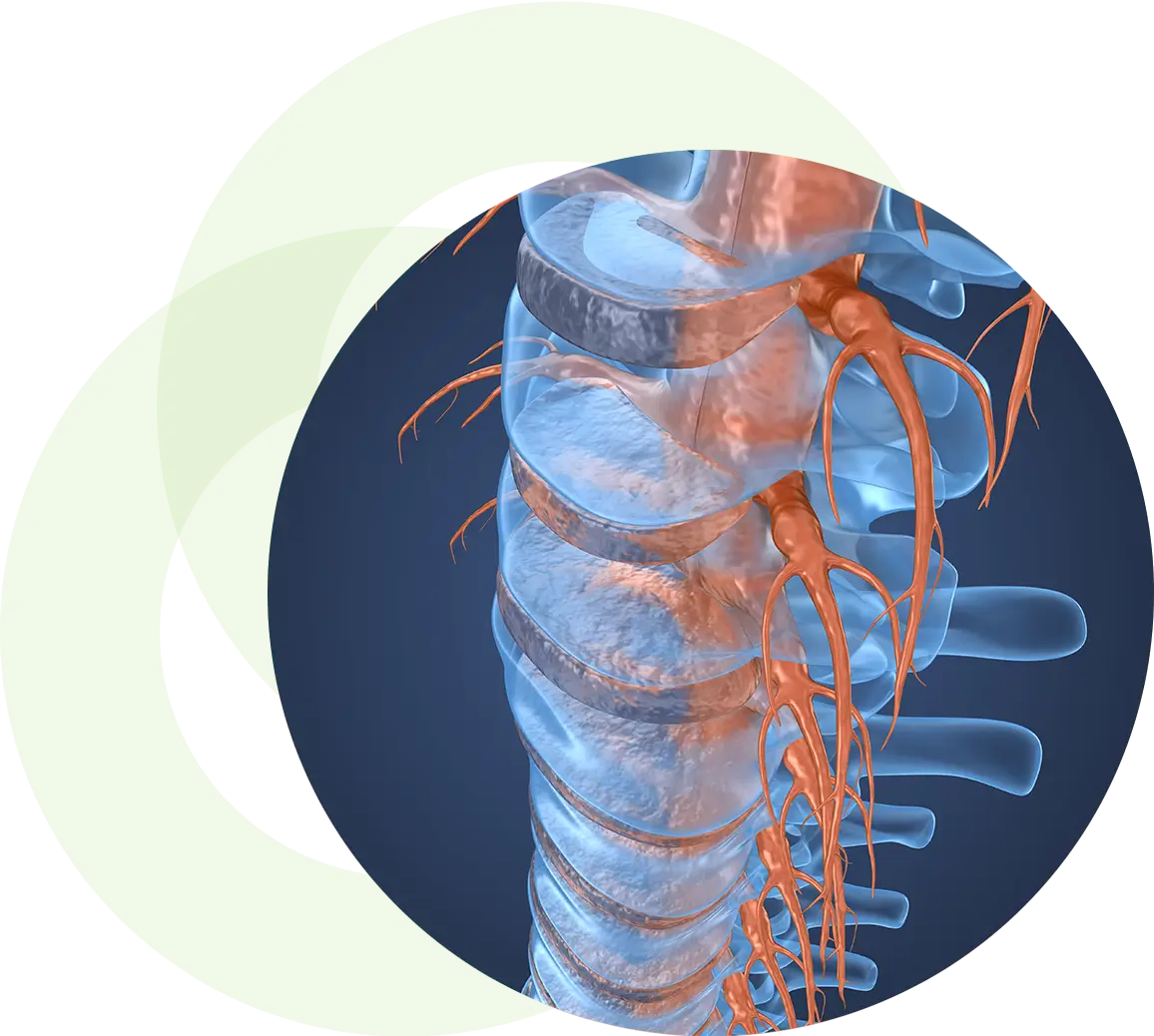
ACDF: Anterior Cervical Discectomy and Fusion
Anterior cervical discectomy and fusion (ACDF) is a type of neck surgery that involves removing a damaged disc to relieve spinal cord or nerve root pressure and alleviate corresponding pain, weakness, numbness, and tingling. A discectomy is a form of surgical decompression, so the procedure may also be called an anterior cervical decompression.
Frequently Asked Questions
The general procedure for an anterior cervical discectomy and fusion—or ACDF—surgery includes the following steps:
- Anterior surgical approach
- The skin incision is one to two inches and is made on the left or right-hand side of the neck. The incision is usually made horizontally within a natural skin crease, though occasionally a more vertical incision is used for multilevel cases.
- The thin muscle under the skin is then split in line with the skin incision, and the plane between the sternocleidomastoid muscle and the strap muscles is then entered.
- Next, a plane between the trachea/esophagus and the carotid sheath is entered.
- A thin fascia (flat layers of fibrous tissue) covers the spine (pre-vertebral fascia), and is then dissected away from the disc space. - Disc removal Fluoroscopy provides an x-ray image of the spine during surgery, and is used to confirm that the spine surgeon is at the correct disc level of the spine.
- After the correct disc space has been identified, the disc is then removed by first cutting the outer annulus fibrosis (fibrous ring around the disc) and removing the nucleus pulposus (the soft inner core of the disc).
- With an anterior cervical discectomy, the entire disc is removed. The cartilage end plates on the vertebral bones are also removed to reveal the hard cortical bone underneath. - Cervical Spine Canal Decompression
- Dissection is carried out from the front to back of a ligament called the posterior longitudinal ligament, which lies between the disc and the spinal cord.
- Often this ligament is gently removed to allow access to the spinal canal to remove any disc material that may have extruded through the ligament, which may be contributing to spinal stenosis.
- The uncinate processes—which are portions of the lower vertebral bone on either side that help form the boundaries of the disc—are typically at least partially removed as well. This is usually where osteophytes (bone spurs) need to be removed in order to relieve spinal cord or nerve root compression.
- The dissection is often performed using an operating microscope or magnifying loupes to aid with visualization of the smaller anatomic structures.

